
Fig. S1
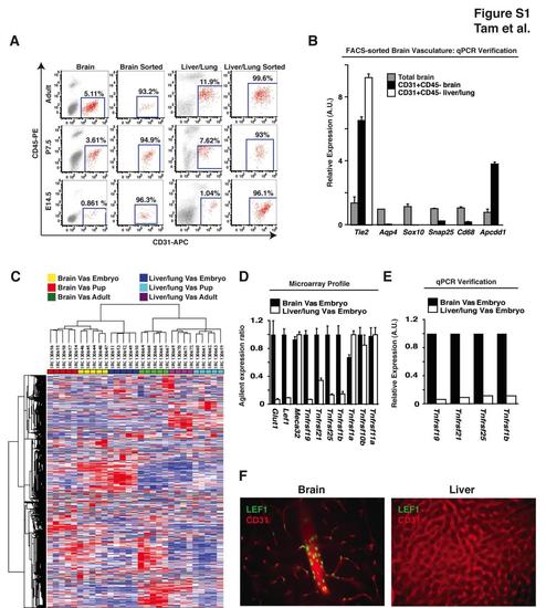
Expression Profiling the Blood-Brain Barrier at Three Developmental Time Points, Related to Figure 1 (A) FACS purification of CD31+CD45- cells from E14.5, P7.5, and adult mice. Single cell suspensions were sorted for CD31+CD45- cells from cerebral cortices and liver/lung tissue to greater than 93% purity. Additional methods were not conducted to further deplete pericytes from the brain vascular cellular preparations. (B) qPCR analysis of FACS purified vasculature. Quantification of transcripts specific to endothelium (Tie2), astrocytes (Aqp4), oligodendrocytes (Sox10), neurons (Snap25), microglia (Cd68), and BBB endothelium (Apcdd1) demonstrate enrichment for BBB endothelium and selection against all other cell types upon FACS purification (Cahoy et al., 2008). (C) Heatmap and dendrogram of microarray expression data from brain and liver/lung tissue. Samples are indicated horizontally across the top of the figure and probes are indicated vertically along the side of the figure. Expression data from the same tissue and developmental time points cluster together, suggesting that these samples are more similar to each other than to the other groups. The following abbreviation is used: Vas, vasculature. (D) Embryonic expression of BBB markers and death receptors. E14.5 embryonic microarray expression data from Figures 1C-E were consolidated. (E) qPCR verification of death receptor enrichment at the BBB. (F) Immunohistochemical analysis of LEF1 expression in the adult mouse cortex and liver. Consistent with our expression profiling data (Figure 1C), we find LEF1-positive nuclei (green) in endothelial cells (CD31: red) from cerebral cortex but not in liver. Results representative of at least three independent experiments are shown. |
Reprinted from Developmental Cell, 22(2), Tam, S.J., Richmond, D.L., Kaminker, J.S., Modrusan, Z., Martin-McNulty, B., Cao, T.C., Weimer, R.M., Carano, R.A., van Bruggen, N., and Watts, R.J., Death Receptors DR6 and TROY Regulate Brain Vascular Development, 403-417, Copyright (2012) with permission from Elsevier. Full text @ Dev. Cell