
Fig. 7
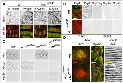
Normal cardiogenesis and myofibrillogenesis in the absence of all Xirps. (A) Neither cardiac nor skeletal muscle sarcomeric organization of myofibrils is affected in 48 hpf xirp1sa0046 mutants. Green: α-Actinin or Myosin; red: Actin. Red scale bars: 10 μm, white scale bars: 5 μm. (B) Sectioned adult cardiac tissue reveals that neither Xirp2a nor Xirp2b are expressed to compensate for the loss of Xirp1. Also, the xirp1sa0046 mutant lacks all three Xirp1 isoforms. Therefore, Xirps are not required for development or maintenance of cardiac tissue. Green: Xirp1; red: Actin. Scale bar: 10 μm. (C) Complete absence of all Xirps within skeletal muscle prior to 24 hpf in xirp1sa0046/xirp2aMO mutant/morphants. Localization of Xirp1 is not affected in xirp2a morphants and, conversely, Xirp2a localization is normal in xirp1sa0046 mutants. Scale bar: 50 μm. (D) Complete loss of Xirps in xirp1sa0046/xirp2aMO mutant/morphants does not impair early cardiogenesis (at 21 hpf) or myofibrillogenesis (at 32 hpf). Green: Xirp1 or Myosin; red: Actin. Red scale bar: 50 μm, white scale bar: 5 μm, yellow scale bar: 20 μm. |
| Genes: | |
|---|---|
| Fish: | |
| Anatomical Term: | |
| Stage: | Long-pec |
| Fish: | |
|---|---|
| Knockdown Reagent: | |
| Observed In: | |
| Stage Range: | 20-25 somites to Long-pec |