
FIGURE
Fig. 3
Fig. 3
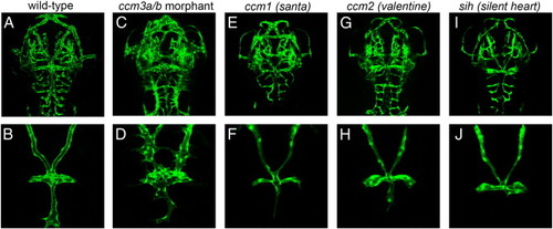
The ccm3a/b cranial vasculature phenotype is specific to ccm3 function and not due to lack of blood flow. Neither santa or valentine mutant embryos display severe vascular dilations or mispatterning seen upon morpholino mediated ccm3a/b knockdown (for san, n = 21, for vtn, n = 32) (E-H) sih embryos similarly displayed no dilations in their cranial vasculature (n = 25) (I and J). Deep confocal z-stack projections (A, C, E, G and I) or z-stacks of dorsal views of the cranial vasculature (B, D, F, H and J) are shown. |
Expression Data
| Gene: | |
|---|---|
| Fish: | |
| Knockdown Reagent: | |
| Anatomical Term: | |
| Stage: | Long-pec |
Expression Detail
Antibody Labeling
Phenotype Data
| Fish: | |
|---|---|
| Knockdown Reagent: | |
| Observed In: | |
| Stage: | Long-pec |
Phenotype Detail
Acknowledgments
This image is the copyrighted work of the attributed author or publisher, and
ZFIN has permission only to display this image to its users.
Additional permissions should be obtained from the applicable author or publisher of the image.
Reprinted from Developmental Biology, 362(2), Yoruk, B., Gillers, B.S., Chi, N.C., and Scott, I.C., Ccm3 functions in a manner distinct from Ccm1 and Ccm2 in a zebrafish model of CCM vascular disease, 121-131, Copyright (2012) with permission from Elsevier. Full text @ Dev. Biol.