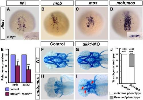
Expression of dkk1, a Wnt signaling inhibitor, is increased in mob;mos embryos, whereas expression of canonical Wnt targets is suppressed. (A–D) Dorsal view of embryos stained with dkk1 riboprobes in wild-type (WT) (A), mob (B), mos (C), and mob;mos (D) at 75% epiboly (8 hpf). dkk1-positive cells are slightly more in mob or mos, but are highly increased in mob;mos embryos. Insets in A and D show transverse sections through the dkk1-expressing region in the mesendoderm. (E) Quantitative PCR analysis of canonical Wnt signaling targets axin2 and sp5l mRNA levels in wild-type embryos and tfap2a-and foxd3-MO knockdown morphants at 8 hpf. Error bars represent standard deviation. Data analyzed by Student′s t-test, **, P < 0.01 and ***, P < 0.001. (F–I) Morpholino-mediated knockdown of dkk1 partially restores pharyngeal arch formation in mob;mos embryos. Ventral view of Alcian blue-stained heads at 5 dpf showing the craniofacial skeleton of wild-type (F), dkk1 morphants (G), mob;mos (H), and dkk1-MO knockdown mob;mos (I) embryos. Head cartilage in mob;mos embryos injected with dkk1-MO is partially restored (red arrows). (J) Percentage of mob;mos embryos with no pharyngeal skeleton and rescued pharyngeal arches with or without (Control) dkk1-MO injection. n indicates the number of embryos examined, N the number of experiments performed. Data analyzed by Chi-squared test; ***, p < 0.001.
|
