
FIGURE
Fig. S2
Fig. S2
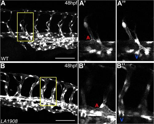
ISVs of both arterial and venous origin are present in LA1908 mutant embryos. A–B, Confocal z-stack projection images of Tg[kdrl:GFP]LA1908 wildtype (A) or mutant (B) embryos. A′–B′′, Single 2.75 μm z-slices of the region outlined in yellow in (A, B), revealing the axial vessel origin of ISVs in day 2 embryos. A′–B′, ISVs originating from the DA are indicated by a red "A". A′′–B′′, ISVs originating from the PCV are indicated by a blue ′′V′′. Scale bars are 100 μm. |
Expression Data
| Gene: | |
|---|---|
| Fish: | |
| Anatomical Term: | |
| Stage: | Long-pec |
Expression Detail
Antibody Labeling
Phenotype Data
| Fish: | |
|---|---|
| Observed In: | |
| Stage: | Long-pec |
Phenotype Detail
Acknowledgments
This image is the copyrighted work of the attributed author or publisher, and
ZFIN has permission only to display this image to its users.
Additional permissions should be obtained from the applicable author or publisher of the image.
Full text @ PLoS One