
Fig. 5
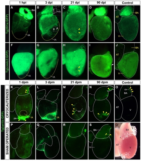
Cardiac tissue regeneration upon cryocauterization. (A-D,F-I) Whole-mount views of dissected hearts of Tg(fli1a:GFP) zebrafish (indicating endocardium and vascular endothelium; A-D) and Tg(cmlc2:GFP) zebrafish (indicating myocardium; F-I) at the indicated times post-injury (hpi or dpi). (E,J) Untreated controls. Anterior is towards the top, ventral towards the right. Broken yellow lines demark the injured area (IA) or the nonregenerated remnant of the IA. (A) Upon cryoinjury, the coronary vasculature and endocardium are completely devoid of GFP expression. (B) At 3 dpi, the IA becomes vascularized (indicated by arrowheads and highlighted in inset box). (C) At 21 dpi, the IA (yellow arrowheads) is nearly indistinguishable from surrounding tissue. (D) By 90 dpi, GFP expression in cryoinjured hearts is similar to control Tg(fli1a:GFP) hearts. (F,G) Lack of myocardial GFP expression in the IA after cryocauterization. (H) Recovery of myocardial GFP expression at 21 dpi; note the small GFP-negative region in the myocardium (yellow arrowheads), possibly a remnant of the IA. (I) By 90 dpi, myocardial GFP expression in cryoinjured hearts is similar to that in control Tg(cmlc2:GFP) hearts. The arrow and broken lines in D and I indicate major long-term morphological alterations in ventricular shape after injury, compared with controls (E,J). (K-S) Whole-mount views of dissected hearts of Tg(wt1b:GFP) zebrafish (indicating epicardium) in cryocauterized hearts (K-N), control hearts (O,O2) and sham-operated hearts (P-S). Yellow arrowheads mark sites of GFP expression. (K) Upon injury, GFP expression is activated on the surface of the heart and pericardium. (L) Expression remains high at 3 dpi, especially close to the IA. (M,N) GFP expression is downregulated at later stages but is still visible at the borders of the IA and in association with coronary vessels. (O) Non-injured control heart revealing just a few GFP-positive cells in the bulbus arteriosus. (O2) Bright-field image of the same heart as in O. (P) Sham-operated heart revealing GFP expression in the epicardium. (Q-S) At 3 dpm, GFP expression is already downregulated and remains low at all later stages analyzed. AT, atrium; BA, bulbus arteriosus; cv, coronary vessel; IA, injured area; V, ventricle. |