
Fig. 1
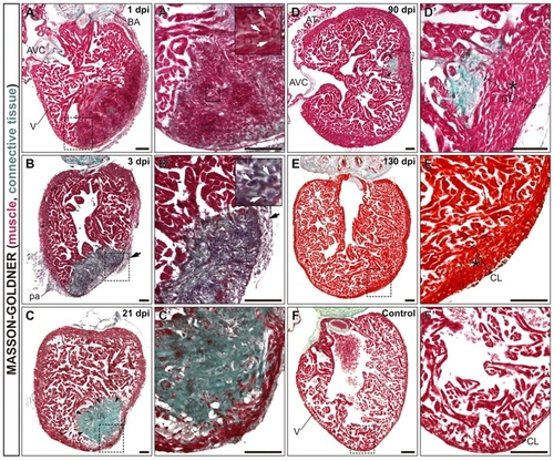
Complete regeneration and scar removal after cryoinjury of the adult zebrafish ventricle. Masson-Goldner-Trichrome stained sagittal sections of adult zebrafish heart fixed at the indicated days after cryocauterization of 25% of the ventricular apex. Anterior is towards the top, ventral towards the right. Boxed areas of the cauterized region are shown at higher magnification in A2-F2. The staining reveals deposition of connective tissue and fibrotic areas in green and healthy myocardial tissue in red. (A,A2) At 1 dpi, there is still some trabeculated myocardium in the injured area (IA). Erythrocytes accumulate at the IA (white arrows in inset). (B,B2) Extensive fibrosis is visible at the IA at 3 dpi. The myocardium has been degraded by this stage. Note the infiltration of the IA with inflammatory cells (white arrows in inset). Black arrows in B and B2 indicate the thickened epicardial layer at 3 dpi. (C,C2) At 21 dpi the compact layer has regenerated and the IA is positioned at a more luminal region. (D,D2) Only a small remnant of the IA is visible at 90 dpi, positioned at the border between compact and trabeculated layers. (E,E2) At 130 dpi, regeneration is complete. Note the enlarged myocardial compact layer near the injury site at 90 dpi (D,D2, asterisk) and 130 dpi (E,E2, asterisk) compared with the control situation (F,F2). Arrows in all panels indicate fibrotic tissue accumulation. AT, atrium; AVC, atrioventricular channel; BA, bulbus arteriosus; CL, compact layer; dpi, days post-injury; IA, injured area; pa, pericardial adhesions; V, ventricle. Scale bars: 100 μm. |