Fig. 3
|
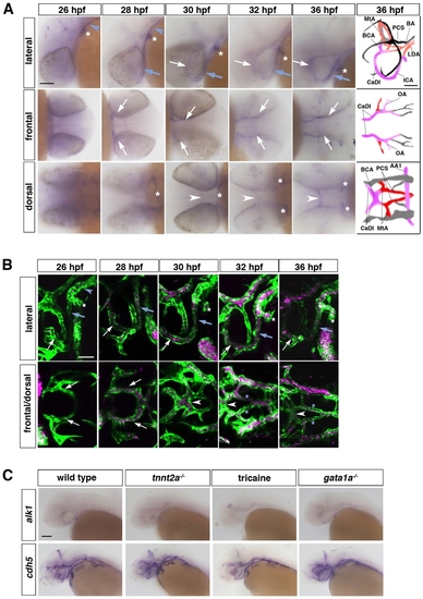
alk1 expression requires blood flow. (A) Spatiotemporal pattern of alk1 mRNA expression assayed by whole-mount in situ hybridization. Expression is detected in the first aortic arch (AA1, white asterisk), lateral dorsal aorta (LDA; blue arrowhead) and internal carotid artery (ICA, blue arrow) at 26-28 hpf, then in the caudal division of the internal carotid artery (CaDI, white arrow) and basal communicating artery (BCA, white arrowhead) by 28-30 hpf. Tracings in the far right column represent all vessels expressing cdh5 at 36 hpf, with alk1-positive arteries in pink, alk1-negative arteries in red and veins in black or gray. BA, basilar artery; LDA, lateral dorsal aortae; MtA, metencephalic artery; OA, optic artery; PCS, posterior communicating segments. Lateral and dorsal views, anterior leftwards. Frontal view, anterior rightwards. Scale bar: 50 μm. (B) Onset of blood flow correlates with alk1 expression. At 26 hpf, blood flows caudally through AA1 (asterisk) and the LDA (blue arrowhead). The cranialward ICA (blue arrow) and CaDI (white arrow) carry flow by 28 hpf; the BCA (white arrowhead) by 30 hpf; and the PCSs (blue asterisks) by 32 hpf. Images are two-dimensional confocal projections of chrna1-/- (paralyzed), Tg(kdrl:GFP)la116;Tg(gata1:dsRed)sd2 embryos. Endothelial cells are green, erythrocytes are magenta. Lateral and dorsal views (row 2, columns 3-5), anterior leftwards. Frontal view (row 2, columns 1-2), anterior rightwards. Scale bar: 50 μm. (C) Whole-mount in situ hybridization demonstrates that alk1 is downregulated in the absence of blood flow (tnnt2a-/- or tricaine treatment, 32-40 hpf) but is not affected by the absence of erythrocytes (gata1a-/-). cdh5 expression is unchanged under all conditions. All embryos are at 36 hpf except tricaine treated, which are at 40 hpf. Lateral views, anterior leftwards. Scale bar: 100 μm. |
| Genes: | |
|---|---|
| Fish: | |
| Condition: | |
| Anatomical Terms: | |
| Stage Range: | Prim-5 to Prim-25 |

