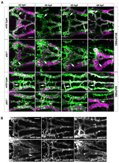
Retention of normally transient arteriovenous connections in alk1 mutants is flow dependent. (A) In wild-type embryos (row 1), transient connections between the basal communicating artery (BCA) and primordial midbrain channel (PMBC) carry blood at 32 hpf but regress by 48 hpf (arrows). In alk1 mutants (row two), one or both of these bilateral connections may be retained, forming a BCA-to-PMBC AVM (arrows). More posteriorly, lumenized connections drain the basilar artery (BA) to the primordial hindbrain channel (PHBC) in wild-type embryos at early times, but almost all regress by 48 hpf (row 3, arrows). In alk1 mutants, one or more of these connections may be retained, forming a BA-to-PHBC AVM (row 4, arrows). Images are two-dimensional confocal projections of Tg(kdrl:GFP)la116; Tg(gata1:dsRed)sd2 embryos, dorsal views, anterior leftwards. Endothelial cells are green; erythrocytes are magenta. (B) AVMs (arrows) are detectable in 48 hpf alk1-/- embryos and in alk1-/-;gata1a-/- embryos (which lack erythrocytes), but not in alk1-/-;tnnt2 MO (which lack blood flow). Images are two-dimensional confocal projections of Tg(fli1a.ep:mRFP-F)pt505 embryos, dorsal views, anterior leftwards. Scale bars: 50 μm.
|

