|
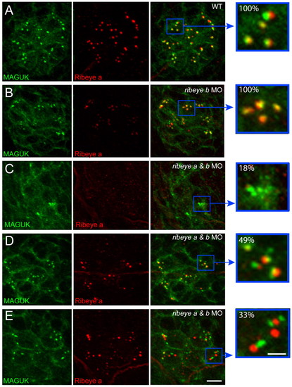
Localization of the postsynaptic density in ribeye morphants. (A-E) Representative confocal z-projections of pan-MAGUK (green) and Ribeye a (red) immunolabel in posterior lateral line neuromast 2 of 4 dpf larvae. Scale bars: 3 μm (main panels), 1 μm (far right panels). (A,B) MAGUK immunolabel juxtaposes Ribeye-labeled punctae in wild-type (A; n=24) and ribeye b MO-injected larvae (B; n=15). Yellow pixels present in the far right panels indicate the close proximity of Ribeye to MAGUK immunolabel. The brightness of Ribeye a immunolabel in B was increased in the merged panel to better visualize its localization. (C) Ribeye immunolabel was absent in 18% (n=33) of ribeye a and ribeye b MO-injected larvae. (D) MAGUK immunolabel appeared to overlap Ribeye in 49% (n=33) of ribeye a and ribeye b MO-injected larvae. (E) MAGUK immunolabel did not overlap Ribeye in 33% (n=33) of ribeye a and ribeye b MO-injected larvae. Yellow pixels are reduced in the far right panel.
|