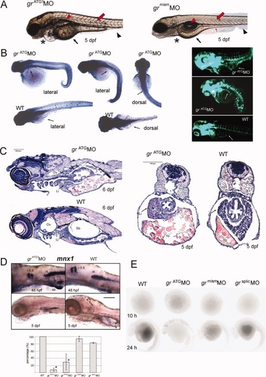
A:grATG1MO-injected embryos failed to inflate the swim bladder (red arrowhead) and displayed a voluminous yolk sac and yolk extension (small arrow), pericardial oedema (asterisk), disrupted myoseptal melanophores (red arrow), and less developed trunk fin (black arrowhead) compared with grmismMO control at 5 dpf. B: Left: alkaline phosphatase staining showing reduced or absent sub-intestinal veins (SIV, arrow) in grATGMO embryos as compared to well-organized SIV in WT embryos at 3 dpf; results were the same using the Tg(fli1a:EGFP)y1 zebrafish transgenic line (right). C: Histology of zebrafish grATG1MO and WT embryos at 6 (sagital sections) and 5 (trasversal sections) dpf. Ov, otic vesicle; Li, liver; In, intestine; Yo, yolk sac; Sb, swim bladder. D: Top: expression of mnx1 in grATG1MO and WT embryos as detected by WISH. Lateral view shows mnx1 expression in the swim bladder epithelium (sb) at 48 hpf and 5 dpf. Other domains of expression are rhombomeres 5 and 6 (r-5,6), neural tube (nt), and endocrine pancreas (p). Bottom: Effects of grATG1MO and grATG2MO, grmismMO, and grsplicMO on swim bladder inflation in larvae at 5 dpf. Results show the percentage average (means ± SD) of individual larvae (n = 20; in triplicate) that developed the gas bladder. Means with asterisks indicate statistically significant differences from controls (P < 0.05). E: TUNEL analysis to detect apoptotic nuclei in WT, grATG1MO grmismMO and grsplicMO embryos at 10 and 24 hpf.
|
