
FIGURE
Fig. 3
- ID
- ZDB-FIG-110321-11
- Publication
- Campos et al., 2011 - Labelling cell structures and tracking cell lineage in zebrafish using SNAP-tag
- Other Figures
- All Figure Page
- Back to All Figure Page
Fig. 3
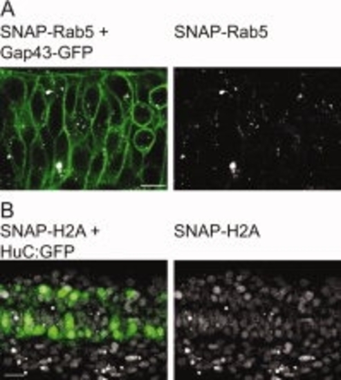
Imaging of SNAP labelled structures is compatible with the use of other FPs. A: Twelve-somite-old embryo co-injected with SNAP-Rab5, BG-Cy3, and Gap43-GFP; single plane confocal micrograph of an embryonic spinal cord (laser power to image SNAP-Rab5: 3%). B: Twenty-two-somite-old transgenic Hu:GFP embryo injected with H2A-SNAP and BG-Cy5 (laser power to image H2A-SNAP: 2%). Projection of 5 planes of the embryonic spinal cord. A,B: Anterior to the left. Scale bars in A,B = 10 μm. |
Expression Data
Expression Detail
Antibody Labeling
Phenotype Data
Phenotype Detail
Acknowledgments
This image is the copyrighted work of the attributed author or publisher, and
ZFIN has permission only to display this image to its users.
Additional permissions should be obtained from the applicable author or publisher of the image.
Full text @ Dev. Dyn.