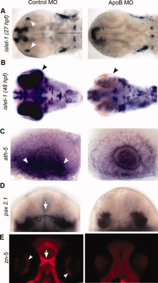
Knockdown of zfApoB phenocopies FucT8 morphants. A,B:islet-1 expression in the optic field is severely reduced (arrowheads) in ApoB morphants, similar to that seen in FucT8 morphants (see Fig. 4A) (27 hpf: 86% of 50 injected embryos showed this phenotype; 48 hpf: 85% of 40 injected embryos showed this phenotype). C: Whole eyes, dissected out from ath-5-labeled embryos, shows a reduced number of ath-5-expressing cells (arrowheads) in 27-hpf ApoB morphants, as seen in FucT8 morphants (see Fig. 4B). Eighty-six percent of 43 injected embryos showed this phenotype. D:pax 2.1 expression in 27-hpf ApoB morphants illustrates a shorter and thicker optic stalk, relative to controls (arrow), and parallels what is seen in FucT8 morphants (see Fig. 4C). Eighty-two percent of 49 injected embryos showed this phenotype. E: Ventral views, anterior to the top. Immunostaining with zn-5 antibody shows loss of RGCs (arrowheads) and optic chiasma (arrow) in 48-hpf ApoB morphant embryos phenocopying that seen in FucT8 morphants (see Fig. 3C). Ninety-eight percent of 47 injected embryos showed this phenotype.
|
