
Fig. S4
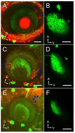
Donor-derived clone location and position-dependent rescue of the retinotectal projection in WT-> radars327 chimeras. (A, C, and E) Lateral confocal projection of the eye. Donor-derived clones were labeled with rhodamine-dextran (blue arrowheads). GFP was only expressed in RGCs of the host. Dorsal up, temporal to the right. (Scale bars, 50μm.) (B, D, and F) Corresponding dorsal confocal projections of tectum. Anterior up, ventral to the right. (Scale bars, 50μm.) (A and B) Donor-derived clones in dorsal eye drive substantial rescue of ventral tectum innervation. (C and D) Donor-derived clones in the ventral eye fail to rescue ventral tectum innervation. (E and F) Donor-derived clones outside of the eye do not rescue ventral tectum innervation. |