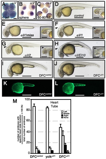
Integrin αV mRNA is a maternal factor and its specific knockdown in DFCs alters heart tube asymmetry. (A-C) WISH analysis in wild-type zebrafish embryos shows maternal expression of αV at the 4-cell stage (A) and sphere stage (4 hpf; B), and zygotic expression at 60-80% epiboly (E). (D-L) Lateral views of live embryos at 32 hpf (D-H) or 28 hpf (I-L). When integrin αV morpholinos (MOs) were delivered at the 1- to 4-cell stage, morphants developed hydrocephaly in the fourth ventricle (black arrowheads) that was also associated with formation of abnormal cerebellum. Control (D) and αV1miss (E) morphants had wild-type head phenotype. Insets in panels D to H represent ∼55 hpf head phenotype of respective morphants. Note intracerebral bleeding (pink color behind eyes) at 55 hpf. When control or αV1 MOs were injected into yolk at mid-blastula stage (512-1000 cells), which targets DFCs specifically, these animals had wild-type phenotype (I,J). (K,L) Fluorescence images corresponding to I and J, revealing that MOs were exclusively present in the yolk cell. (M) Bar graph showing the effects of αV integrin loss-of-function specifically in DFCs on heart tube location. Data expressed are similar to those in Fig. 1E. Scale bars: 100 μm in A-C; 500 μm in D-L. See also Table S3 in the supplementary material.
|
