
Fig. S1
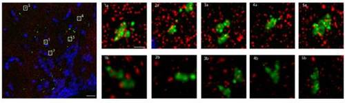
The presynaptic marker SYP-EGFP colocalizes with the post synaptic density 95 (PSD95) protein. Array tomography assay. Large Panel: Composite max projections of brain tissue from the hypothalamus of hcrt:SYPEGFP fish in a volumetric section 107.2μm by 107.4μm by 2.1μm. Red = PSD95, Green = SYP-EGFP, Blue = DAPI. Scale bar = 10μm. Small Panels: Five representative selections of SYP-EGFP puncta. 1a, 2a, 3a, 4a, 5a are max projections of 4.1μm by 4.1μm by 2.1μm subsections of the larger volume. Note the degree of superposition of PSD95 (red) to SYP-EGFP (green). Scale bar = 1μm. 1b, 2b, 3b, 4b, 5b are 3 dimensional renderings of the same piece of tissue. Note that many of the PSD95 (red) puncta that superimpose on SYP-GFP (green) are definitely separated in 3D space. It is clear, however, that for each SYP-EGFP punctum there are smaller corresponding PSD95 puncta that reside in close juxtaposition with the presynaptic marker. Out of 25 randomly selected puncta, 21 had at least 1 punctum of PSD95 within 100nm of the SYP-EGFP puncta. |