
FIGURE
Fig. S3
- ID
- ZDB-FIG-101013-7
- Publication
- Van Otterloo et al., 2010 - Differentiation of zebrafish melanophores depends on transcription factors AP2 alpha and AP2 epsilon
- Other Figures
- All Figure Page
- Back to All Figure Page
Fig. S3
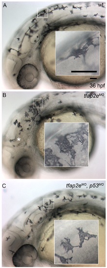
p53 MO blocks nervous system necrosis but does not affect melanophore development. Lateral views of live zebrafish embryos at 36 hpf. Insets show higher magnification of melanophores contained in white boxes. (A) A wild-type embryo shows normal melanophore development, similar to embryos injected with tfap2e e3i3 MO (B,C). (B) The embryo injected with tfap2e MO also displays signs of central nervous system cell death (i.e., patches of opacity in the brain and spinal cord, white asterisk), which is reversed (C) by co-injection of a p53 MO. |
Expression Data
Expression Detail
Antibody Labeling
Phenotype Data
Phenotype Detail
Acknowledgments
This image is the copyrighted work of the attributed author or publisher, and
ZFIN has permission only to display this image to its users.
Additional permissions should be obtained from the applicable author or publisher of the image.
Full text @ PLoS Genet.