|
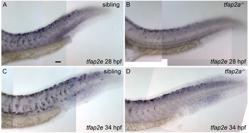
Expression of tfap2e in tfap2a mutants. Lateral views of zebrafish embryos, fixed at the stage indicated and processed to reveal tfap2e expression by RNA in situ hybridization. (A) A sibling embryo at 28 hpf with tfap2e expression within melanoblasts, located throughout the trunk of the embryo, as described earlier. (B) A tfap2a mutant, in which tfap2e expression is detected within melanoblasts near the dorsum of the embryo; it is evident that fewer than normal numbers of tfap2e-expressing cells (presumed melanoblasts) have migrated ventrally. (C) Sibling and D) tfap2a mutant embryos at 34 hpf; tfap2e expression is detected in the posterior trunk of both sibling and mutant embryos, although fewer tfap2e-expressing cells have migrated ventrally in the tfap2a mutant. Embryos were treated with low levels of PTU to better visualize expression within melanophores. Scale bar: 25 μM.
|