
Fig. 3
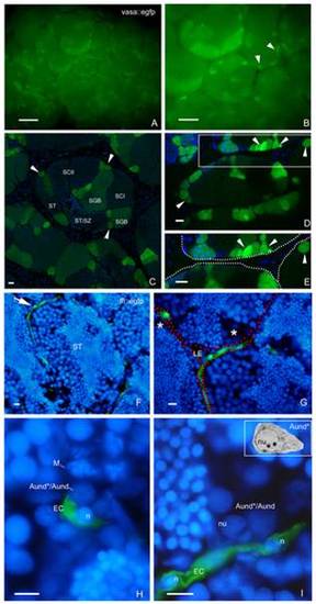
SSC niche in zebrafish testes. Whole-mount of vasa::egfp testes analyzed under fluorescence (A,B) and CLSM (C–E). A,B. Most of the brightest and small spots (type A undifferentiated spermatogonia) (arrowheads) are adjacent the triangle/lozenge dark areas (interstitium). Scale bars = 0.1 mm and 0.5 mm, respectively. C–E. Vasa is highly expressed in type A undifferentiated spermatogonia, and is gradually decreased during the spermatogenesis. Aund*/Aund (arrowheads), type B spermatogonia (SGB), primary spermatocytes (SCI), secondary spermatocytes (SCII), spermatids (ST), spermatids and spermatozoa (ST/SZ). Note that most of Aund*/Aund (arrowheads) are situated near the interstitium (D, dark areas; E, delimited by spotted lines). E is a high magnification of the square in D. Scale bars = 10 μm. F–I: Cryosections of fli::egfp testes stained with DAPI (nuclear staining) and analyzed under fluorescence microscopy. The arrow in F shows a blood vessel (green) surrounding the circumference of a seminiferous tubule (ST). Interstitium (delimited with red dotted lines), Leydig cells (LE) and group of type A spermatogonia (asterisks) are shown in G. H,I. Aund*/Aund are near the endothelial cell (EC) nuclei (n). Note a metaphase figure (M) in H. Nucleolus (nu) of type A undifferentiated spermatogonia is shown in I. Compare the similar morphology of Aund* (inset) with the cells found near the endothelial cell. Scale bars = 10 μm. |
| Gene: | |
|---|---|
| Fish: | |
| Anatomical Terms: | |
| Stage: | Adult |