
Fig. 4
- ID
- ZDB-FIG-100816-16
- Publication
- Goody et al., 2010 - Nrk2b-mediated NAD+ production regulates cell adhesion and is required for muscle morphogenesis in vivo: Nrk2b and NAD+ in muscle morphogenesis
- Other Figures
- All Figure Page
- Back to All Figure Page
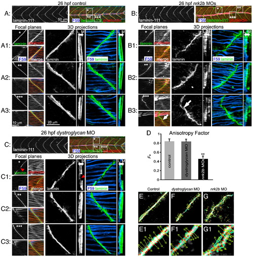
Nrk2b is required for normal basement membrane development. (A, B, C) 26 hpf embryos, side mount, anterior left, dorsal top, F59 antibody stained to visualize slow-twitch muscle, phalloidin stained to visualize actin and Laminin-111 antibody stained to visualize the basement membrane of the MTJ. Also see Supplementary movies. (A) Control. (B) nrk2b morphant. (C) dystroglycan morphant. Numbered panels 1–3 are examples of different MTJs within an embryo from each treatment (*, ** or *** indicates the location of the MTJ). Small square panels are focal planes of each antibody/stain used as well as a merged view. Large square panels are 3D reconstructed Laminin-111 antibody staining (in black and white) or F59 and Laminin-111 antibody staining (in color). Tall rectangular panels are transverse views of the 3D reconstructed antibody staining with lateral to the left and medial to the right. Note in the MTJ examples in nrk2b morphants (panel B) that Laminin-111 is not laid down in a straight line and is not always continuous (white arrows). In the transverse views, note that Laminin-111 is especially disrupted in the medial fast-twitch fiber domain. In dystroglycan morphants, MTJs can still be discontinuous (red arrowheads), but Laminin-111 is robust in the lateral and medial domains. (D) Graph of the anisotrophy factor (see Materials and methods) in Laminin-111 stained images. A lower anisotrophy value in nrk2b morphant images indicates that there is less order/structure in Laminin-111 staining in these morphants. (E–G) Side mount, anterior left, dorsal top, 26 hpf embryos, Laminin-111 antibody staining. Numbered panels are higher magnification views of corresponding panels. Images were analyzed with the 2D Wavelet Transform Modulus Maxima (2DWTMM) method. (Panel E) Controls. (Panel F) dystroglycan morphants. (Panel G) nrk2b morphants. In controls and dystroglycan morphants, the vectors are more parallel indicating more order than in nrk2b morphants. Scale bars are labeled on the figure, **p < 0.01. |
| Fish: | |
|---|---|
| Knockdown Reagents: | |
| Observed In: | |
| Stage: | Prim-5 |
Reprinted from Developmental Biology, 344(2), Goody, M.F., Kelly, M.W., Lessard, K.N., Khalil, A., and Henry, C.A., Nrk2b-mediated NAD+ production regulates cell adhesion and is required for muscle morphogenesis in vivo: Nrk2b and NAD+ in muscle morphogenesis, 809-826, Copyright (2010) with permission from Elsevier. Full text @ Dev. Biol.