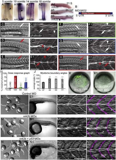
nrk2b expression and MO characterization. (A–A6) ISH with nrk2b probe. (A–A3) Dorsal mount, anterior top. (A4–A6) Side mount, anterior left, dorsal top. Black arrowheads denote nrk2b expression. (B) Cartoon of nrk2b and MO target sites. (C–H1) Side mount, anterior left, dorsal top, 26 hpf embryos, lettered panels are DIC images, numbered panels are phalloidin stained to visualize actin. (C, C1) Injection of a standard control MO did not elicit a phenotype. Myotomes are V-shaped (white arrows) and muscle fibers are normal (white arrowheads). Injection of the functional dose of MO1 (D, D1) or MO2 (E, E1) resulted in U-shaped myotomes (red arrows) and wavy muscle fibers (red arrowheads). Injection of half the functional dose of MO1 (F, F1) or MO2 (G, G1) did not disrupt muscle development. (H, H1) Injection of both low doses of MO1 + MO2 recapitulated the phenotype. (I) Dose response graph. Injection of low doses of MO1 + MO2 resulted in more embryos with the phenotype than injection of either low dose alone (n of embryos injected is listed on the x-axis). (J) Average myotome boundary angles of 26 hpf control, laminin γ1 mutants, nrk2b morphant, and nrk2b + p53 MO-injected embryos. laminin γ1 mutants, nrk2b morphants, and nrk2b + p53 morphants have myotome boundaries of a similar angle and all have myotome boundary angles that are significantly wider than controls, p < 0.001 (n of MTJs measured is listed on the x-axis). (K–K1) Brightfield images of shield stage embryos. Animal pole to the top. (K) Embryo injected with the nrk2b:gfp plasmid that contains both MO target sites. (K1) Embryo injected with the nrk2b:gfp plasmid and nrk2b MOs. Note that expression of GFP-tagged Nrk2b is drastically decreased by injection of nrk2b MOs (3 experiments, 1 representative experiment: 93% of nrk2b:gfp injected embryos expressed GFP (n = 171 out of 184 embryos) whereas only 5% of nrk2b:gfp + nrk2b MO-injected embryos expressed any GFP (n = 1 out of 20 embryos)). (L–N) Brightfield images of a dish of 26 hpf embryos. (L–L1) Control MO-injected embryos. (M–M1) nrk2b morphants. (N–N1) nrk2b + p53 morphants. (L1–N1) Side mount, anterior left, dorsal top, 26 hpf embryos, DIC imaging. Higher magnification images of one representative embryo from each of the corresponding dishes. Note that the control morphant (L1) has a longer body axis than both nrk2b morphants (M1) and nrk2b + p53 double morphants (N1). (L2–N3) Side mount, anterior left, dorsal top, 26 hpf embryos, phalloidin staining to visualize actin. Higher magnification views of embryos from the corresponding dishes. In panels numbered 3, myotome boundaries were pseudocolored fuchsia and the myotome boundary angle is given with the corresponding boundary. Note that while injection of p53 MO did rescue the cell death seen in the head of nrk2b morphants, it did not rescue the shorter body axis (compare M1 to N1) or the U-shaped myotomes (compare M3 to N3). Scale bars are 50 μm, ***p < 0.001.
|
