FIGURE

Fig. 3

ID
ZDB-FIG-100309-48
Publication
Poon et al., 2010 - Zebrafish cardiac enhancer trap lines: New tools for in vivo studies of cardiovascular development and disease
Other Figures
All Figure Page
Back to All Figure Page
Fig. 3

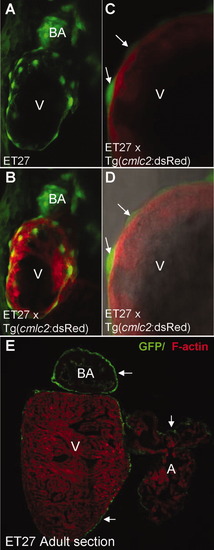
CETs that define the epicardium in vivo. A, B: ET27 with EGFP-positive cells covering the ventricle; note that EGFP expression in the BA precedes that in the epicardium; C, D: The EGFP-positive epicardial cells (arrow) enveloping the internal cmlc2 highlighted myocardium at high magnification. E: Cross-section of the adult ET27 heart at low magnification, with GFP-labelled epicardium on the external surface of the BA, atrium and ventricle (arrow). B-D: Double transgenic embryos of ET27 and Tg(cmlc2:dsRed). All except D are fluorescent images. D: Composite fluorescent/DIC images. A, atrium; BA, bulbus arteriosus; V, ventricle.

Expression Data
Gene:
Fish:
Anatomical Terms:
Stage: Protruding-mouth

Expression Detail
Antibody Labeling
Phenotype Data

Phenotype Detail
Acknowledgments
This image is the copyrighted work of the attributed author or publisher, and ZFIN has permission only to display this image to its users. Additional permissions should be obtained from the applicable author or publisher of the image. Full text @ Dev. Dyn.