|
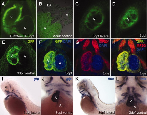
CETs that define development of the myocardium in vivo. A: EGFP-positive embryonic myocardium in the ventricle is thicker compared to that in the atrium. B: EGFP expression in adult heart sections of myocardium-specific ET33-mi3A. C, D: Single optical sections of ventricular myocardium in the live embryo at high magnification (lateral view). E: High-resolution 3D-reconstruction of myocardium live. F-H: Myocardium immunolabelled with EGFP/MF20(red)/DAPI(blue). A, C, D, E: In vivo imaging of live embryos at 3 dpf. I,J: Lateral and ventral view of larva after WISH with anti-gfp probe. K, L: Lateral and frontal view of larva after WISH with anti-fhla probe. All images are ET33-mi3A. A, atrium; BA, bulbus arteriosus; V, ventricle.
|