|
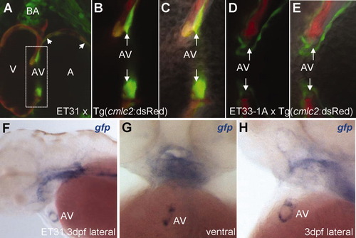
The CET line, ET31, marks a unique subset of myocardium at the early A-V valve. A: ET31 crossed with Tg(cmlc2:dsRed) showing a subset of myocardium positive for EGFP at the A-V junction and chambers (arrowhead). Note the EGFP expression at the BA. B, C: High magnification of A-V junction reveals differential GFP intensity between a different subset of myocardial cells. Arrows point to non-overlapped ET31 EGFP expression. D, E: High magnification of A-V junction lined with endocardial cells adjacent to cmlc2 marked myocardium (shown for comparison). Arrows point to A-V junction that ET31 marks. F-H: anti-gfp WISH demonstrates that the expression domain at the A-V node represents a ring. A-C: Double transgenic embryos of ET31 and Tg(cmlc2:dsRed). D, E, Double transgenic embryos of ET33-1A and Tg(cmlc2:dsRed). B, D: Fluorescent images. C, E: Composite fluorescent/DIC images to reveal cellular morphology. A, atrium; BA, bulbus arteriosus; A-V, atrio-ventricular valve; V, ventricle.
|