FIGURE
Fig. S4
Fig. S4
|
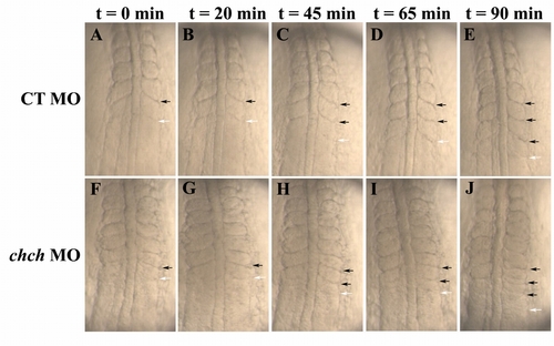
Pace of the "molecular clock" is not significantly altered in ChCh-compromised embryos. Living wild-type and ChCh-compromised embryos beginning at the seven-somite stage. A-J: All views are dorsal, anterior to the top. Duration of somite formation in both control (A-E) and ChCh-compromised embryos (F-J) is approximately 45 min at 23°C. Black arrows denote already formed somite boundaries and white arrows denote newly forming segmentation furrow. |
Expression Data
Expression Detail
Antibody Labeling
Phenotype Data
Phenotype Detail
Acknowledgments
This image is the copyrighted work of the attributed author or publisher, and
ZFIN has permission only to display this image to its users.
Additional permissions should be obtained from the applicable author or publisher of the image.
Full text @ Dev. Dyn.

