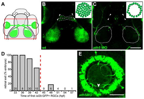
Early RGCs are necessary for axons to exit eye. (A) Diagram showing field of view for B,C. (B,C) 5 dpf isl2b:GFP, confocal z-projections; dorsal views, anterior upwards. Insets show cell bodies in eye. (B) Wild-type axons exit eye (arrowheads), project through the optic chiasm (broken lines) and to the optic tecta. (C) With high dose of ath5 MO, axons do not exit eyes (arrowheads) or innervate tecta (outlined). (D) Retinal exit in dose-response experiment of Fig. 1D, plotting percentage of embryos in which axons exit the eye against the time at which their first isl2b:gfp-positive RGCs were born. When RGCs are born by 42 hpf, axons usually exit the eye; when delayed after 42 hpf, axons rarely exit. Number of embryos is indicated at base of each bar. (E) 72 hpf isl2b:GFP, confocal z-projection; dorsal upwards. In a high-dose ath5 morphant, axons from peripheral RGCs remain trapped in the RGC layer without entering the optic nerve. To better appreciate 3D structure, see volume reconstruction in Movie 1 in the supplementary material. Arrowhead indicates the optic nerve head, stained by anti-Pax2 (not shown). Scale bars: 100 μm in C; 50 μm in E.
|
