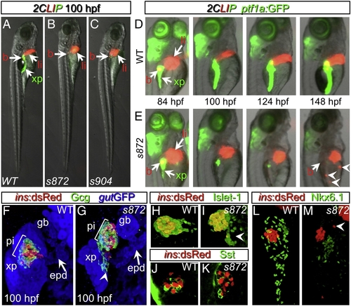
Initial formation and degeneration of the exocrine pancreas in dandelion (ddn) mutants. (A–C) Lateral views of WT (A), ddns872 (B), and ddns904 mutant (C) larvae at 100 hpf in the 2-color Liver, Insulin, acinar Pancreas (2CLIP) transgenic background (see Materials and methods). Acinar cells of the exocrine pancreas (xp) are green and hepatocytes (li) and pancreatic beta cells (b) are red. ddn mutants exhibit minimal acinar tissue, but overall larva morphology is only moderately affected. (D, E) Successive images of individual WT (D) and ddns872 mutant (E) larvae between 84 and 148 hpf in the 2CLIP; Tg(ptf1a:EGFP)jh1 background. The exocrine pancreas and liver grow larger in WT, but degenerate in ddn mutants. Hepatocyte fragments are found in the circulation (arrowheads). (F–K) 3-D projections of confocal stacks showing composition of WT (F, H, J) and ddns872 mutant (G, I, K) endocrine pancreas at 100 hpf in Tg(ins:dsRed)m1081 background. (F, G) A core of Tg(ins:dsRed)m1081+ cells (red) is surrounded by a mantle of Glucagon+ (Gcg) cells (green). Extrapancreatic duct (epd) structure is intact. (H, I) Isl-1, and (J, K) Somatostatin (Sst) positive cells appear unaffected. Arrowheads in G, I, and M point to endocrine cells outside of the primary islet. (L, M) Nkx6.1 immunostaining reveals the presence of intra-pancreatic duct cells in both WT and dnmt1s872 mutant larvae.
|
