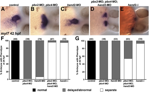
Pbx and Hand2 genetically interact in cardiac morphogenesis. (A–E) RNA in situ expression of myl7 at 42 hpf in (A) control, (B) pbx2-MO; pbx4-MO, (C) hand2-MO, (D) pbx2-MO; pbx4-MO; hand2-MO, or (E) hand2-/- embryos. Embryos are shown in frontal views, dorsal towards the right. (F–G) Quantification of phenotypes observed using myl7 expression at (F) 24 hpf or (G) 42 hpf. Normal phenotypic classes (black) correspond to tube (24 hpf) or looped (42 hpf) patterns depicted in Fig. 4D and subpanel A of this figure, respectively. Delayed or abnormal phenotypic classes (gray) correspond to images shown for pbx2-MO; pbx4-MO and hand2-MO embryos in Fig. 4D (24 hpf) or subpanels B–C of this figure (42 hpf). The separate, or cardia bifida, phenotypic classes (white) correspond to two myl7 domains and resemble those described for pbx2-MO; pbx4-MO and hand2-MO embryos in Fig. 4A or for pbx2-MO; pbx4-MO; hand2-MO and hand2-/- embryos in subpanels D–E of this figure. Numbers in parentheses denote numbers of embryos. hand2-/- embryos were derived from clutches that contained about 25% mutant embryos; their hand2+/+ and +/- siblings showed normal myl7 expression.
|
