
Fig. 7
- ID
- ZDB-FIG-090831-7
- Publication
- Insinna et al., 2009 - Different roles for KIF17 and kinesin II in photoreceptor development and maintenance
- Other Figures
- All Figure Page
- Back to All Figure Page
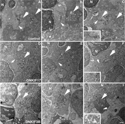
Lack of synaptic ribbons in cone pedicles of DNKIF3B-expressing embryos. A-C: Control pedicles showing postsynaptic invaginations (large arrows) and synaptic ribbons (small arrows) associated with presynaptic membranes. D-F: DNKIF17 cone pedicles showing postsynaptic invaginations (large arrows) and synaptic ribbons (small arrows) associated with presynaptic membranes. G-I: DNKIF3B cone pedicles showing postsynaptic invaginations (large arrows) without synaptic ribbons. Occasionally, ribbons are seen dissociated from presynaptic membranes (small arrow in I). Condensed nuclei and pedicles (asterisks) of dying cells are seen in G. Insets in C, F, and I are enlargements of invaginations to show association of ribbons with the presynaptic membrane. Scale bars = 500 nm in I and in inset. |