
Fig. 6
- ID
- ZDB-FIG-090831-6
- Publication
- Insinna et al., 2009 - Different roles for KIF17 and kinesin II in photoreceptor development and maintenance
- Other Figures
- All Figure Page
- Back to All Figure Page
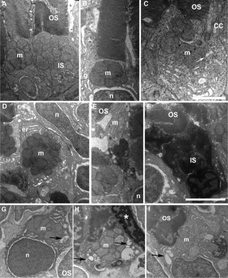
Accumulation of large vacuoles and dense material in the inner segment (IS) of cones expressing DNKIF3B. A: Electron microscopy (EM) view of cones at 5 days after injection of the Ta-CP driving green fluorescent protein (GFP) alone (control). Note the normal structure of outer segment (OS) discs and IS with accumulation of mitochondria (m) in the ellipsoid region. B,C: EM view of cones at 5 days after injection of the Ta-CP driving DNKIF3B showing mild IS defects. This was primarily enlargement of the cytoplasmic area around the mitochondria. Arrow indicates large vesicle below the connecting cilium (CC). n, photoreceptor nucleus; g, Golgi apparatus. D: EM view of a cross-section through the IS of a cone with a mild IS phenotype involving enlargement of the cytoplasmic area around mitochondria; er, endoplasmic reticulum. The upper arrow indicates accumulation of large vesicles in the ellipsoid region. The lower arrow indicates a Golgi region. E: EM view of a cone in the central retina that has a highly condensed IS between the nucleus (n) and mitochondrial rich (m) ellipsoid region. OS discs are normal. F: Cone with a highly condensed nucleus and IS with normal OS organization. G,H: Examples of cones accumulating vesicles and large vacuoles (arrows) within the IS surrounding the mitochondria. Asterisk in H indicates condensed nucleus of an adjacent cone. Scale bar = 1.5 μm in C, 2.5 μm in F. |