
Fig. 3
- ID
- ZDB-FIG-090831-4
- Publication
- Insinna et al., 2009 - Different roles for KIF17 and kinesin II in photoreceptor development and maintenance
- Other Figures
- All Figure Page
- Back to All Figure Page
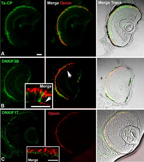
Localization of Ta-CP-directed expression of green fluorescent protein (GFP), DNKIF3B, and DNKIF17 and cone opsin localization at 5 days postfertilization (dpf). The Ta-CP promoter up stream of GFP, DNKIF3B, or DNKIF17 was injected at the one-cell stage. A: In Ta-CP/GFP embryos, GFP fluorescence was seen throughout the outer retina and cone opsin was highly localized to the outer segment (OS). B: In Ta-CP/DNKIF3B embryos, transgene expression (GFP, green) was similar to controls, but cone opsin was mislocalized to the perinuclear region and outer plexiform layer (arrow) across the entire retina. Inset: Higher power image showing cone opsin mislocalization to the synaptic layer (arrow). C: In Ta-CP/DNKIF17 embryos transgene expression (GFP, green) was distributed across the entire retina, but was largely restricted to the inner segment (IS; see inset). Cone opsin was in the OS, and there was no evidence of mislocalization. Inset: Higher power image showing lack of mislocalization of cone opsin and strong accumulation of DNKIF17 in the IS. Scale bars = 10 μm. The bar in A applies to panels A-C. |