
Fig. 4
- ID
- ZDB-FIG-090831-5
- Publication
- Insinna et al., 2009 - Different roles for KIF17 and kinesin II in photoreceptor development and maintenance
- Other Figures
- All Figure Page
- Back to All Figure Page
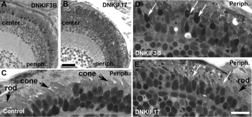
Light microscopy of retinae of control, DNKIF3B, and DNKIF17 embryos at 5 days postfertilization (dpf). A,B: Low power images of semi-thin plastic sections of DNKIF3B (A) and DNKIF17 (B) eyes. Central (center) and peripheral (periph) retinal regions are labeled. C: Higher power image of the outer retina of a control eye; the periphery is on the right (Periph). Cone and rod outer segment (OS) are indicated by arrows. D,E: Higher power images of outer retina of DNKIF3B (D) and DNKIF17 (E) eyes; the periphery is on the right (periph). White arrows in D indicate condensed nuclei and cell bodies of dying cones. White arrows in E indicate cones with very short or missing OS. A normal rod OS is indicated in E. Scale bars = 10 μm. The bar in E applies to C-E. |