
Fig. 1
- ID
- ZDB-FIG-090817-9
- Publication
- McMahon et al., 2009 - Lmx1b is essential for survival of periocular mesenchymal cells and influences Fgf-mediated retinal patterning in zebrafish
- Other Figures
- All Figure Page
- Back to All Figure Page
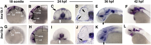
Zebrafish lmx1b.1 and lmx1b.2 are expressed in the ocular and non-ocular tissue. In situ hybridization of lmx1b.1 (A–F) and lmx1b.2 (G–L) from 18 s to 42 hpf. Anterior is to the left in lateral views. Expression of both lmx1b genes was strong in the midbrain–hindbrain boundary and ventral diencephalon at 18 s (A, G) and 24 hpf (B, H). Only lmx1b.1 was detected in the otic vesicle (A versus G) and spinal cord neurons (B versus H, boxed). For both genes, periocular expression in cells associated with the optic stalk (white arrows C, I) and the globe of the eye (black arrows C, I), was observed in whole mount and histological sections (arrows D, J). Expression of lmx1b.1 and lmx1b.2 was also detected in the ventral fissure (arrows E, K) and hindbrain neurons (E, F and K, L). See text for additional details. mhb, midbrain–hindbrain boundary; vd, ventral diencephalon; dd, dorsal diencephalon; ov, otic vesicle; hb, hindbrain. |
| Genes: | |
|---|---|
| Fish: | |
| Anatomical Terms: | |
| Stage Range: | 14-19 somites to High-pec |
Reprinted from Developmental Biology, 332(2), McMahon, C., Gestri, G., Wilson, S.W., and Link, B.A., Lmx1b is essential for survival of periocular mesenchymal cells and influences Fgf-mediated retinal patterning in zebrafish, 287-298, Copyright (2009) with permission from Elsevier. Full text @ Dev. Biol.