
Fig. 7
- ID
- ZDB-FIG-090817-15
- Publication
- McMahon et al., 2009 - Lmx1b is essential for survival of periocular mesenchymal cells and influences Fgf-mediated retinal patterning in zebrafish
- Other Figures
- All Figure Page
- Back to All Figure Page
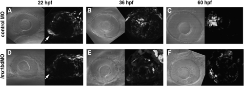
Migration of lmx1b.1:GFP-positive cells is altered following knock-down of Lmx1b. Bright field and fluorescence images of (panels A–C) control MO and (panels D–F) lmx1bdMO embryos from the Tg(- 5 kb lmx1b.1:GFP)mw11 line. For each image developmental time is indicated at the top, anterior is to the left, and dorsal is up. Arrows indicate the location of the optic stalk region. (D) Note the significant reduction of GFP fluorescence in Lmx1b-deficient cells associated with the optic stalk. Each fluorescent image is a compressed z-stack, which includes the entire globe of the eye and optic stalk region. Images shown are from representative embryos (n = 12 total embryos tracked over time from 2 experiments). |
| Gene: | |
|---|---|
| Fish: | |
| Knockdown Reagents: | |
| Anatomical Terms: | |
| Stage Range: | 26+ somites to Pec-fin |
| Fish: | |
|---|---|
| Knockdown Reagents: | |
| Observed In: | |
| Stage Range: | 26+ somites to Pec-fin |
Reprinted from Developmental Biology, 332(2), McMahon, C., Gestri, G., Wilson, S.W., and Link, B.A., Lmx1b is essential for survival of periocular mesenchymal cells and influences Fgf-mediated retinal patterning in zebrafish, 287-298, Copyright (2009) with permission from Elsevier. Full text @ Dev. Biol.