
Fig. S2
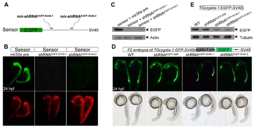
Transient knockdown of chromosomally integrated EGFP gene. (A) Diagram of mir-shRNAEGFP-SV40-1 and mir-shRNAEGFP-SV40-2 against the proximal and distal SV40-3′UTR of EGFP, respectively. (B) Detection of EGFP and DsRed fluorescence in 24 hpf embryos injected with indicted mRNAs. Red fluorescence was used as an injection control. (C) Western blot analysis of 24 hpf embryos shown in panels B. The β-actin was used as a loading control. (D) Detection of EGFP fluorescence in the Tg(zgata-1:EGFP-SV40) transgenic embryos injected with indicated mRNAs. The development and morphology of injected embryos appeared to be normal (bottom panels). (E) Western blot analysis of 24 hpf embryos shown in panels D. The α-tubulin was used as a loading control. |