
Fig. 5
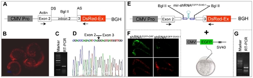
Design of pol II promoter-driven knockdown construct. (A) Diagram of pol II-type promoter CMV driven-knockdown vector (CMV promoter-actin-DsRed-BGH). DS: donor site; AS: acceptor site. The first 21-base pairs of exon 3 of zebrafish actin gene have been in-frame fused to the DsRed fluorescent protein gene followed by a bovine growth hormone (BGH) sequence as 3′UTR. (B) Red fluorescence was observed in 22 hpf embryos injected with the plasmid shown in panel A. (C) RT-PCR analysis with total RNAs derived from 22 hpf embryos shown in panel B. The primers used are indicated by horizontal arrows in panel A. (D) The sequence of RT-PCR product shown in pane C. Note that the entire intron 2 of the actin gene has been spliced out (arrow). (E) The mir-shRNAEGFP-SV40-1 was inserted into the intron 2 at the Bgl II site and co-injection with CMV-EGFP-SV40 reporter plasmid. (F) Knockdown of EGFP fluorescence in the 24 hpf embryos co-injected with EGFP-SV40 reporter plasmid plus CMV promoter-actin-DsRed-BGH plasmid carrying either mir-shRNAEGFP-ORF or mir-shRNAEGFP-SV40-1. (G) RT-PCR analysis of total RNAs derived from 22 hpf embryos shown in panel F. |