
Fig. S1
- ID
- ZDB-FIG-081104-8
- Publication
- Batista et al., 2008 - Pax2/8 act redundantly to specify glycinergic and GABAergic fates of multiple spinal interneurons
- Other Figures
- All Figure Page
- Back to All Figure Page
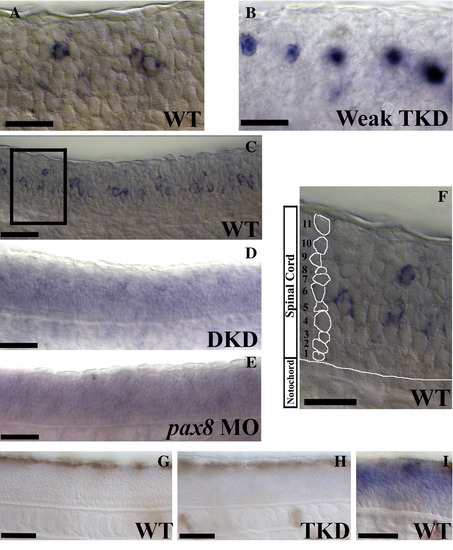
Lateral views of pax8 expression in 24 h trunks (A–F) and p53 expression in 36 h trunks (G–I). pax8 morpholinos block the splicing of pax8 RNA causing it to be retained in the nucleus (see discussion of splice-blocking morpholinos in Yan et al., 2002). (A) Cytoplasmic pax8 RNA in wild-type embryos; (B) unspliced nuclear pax8 RNA in noi (pax2a) mutants injected with 1 mg/ml each of pax2b and pax8 MOs (“weak” triple knock-down (TKD) embryos). When these morpholinos are injected at a higher concentration, expression of pax8 is lost (see Fig. 1F in the main paper). pax8 expression depends at least partly on Pax2 function. In noi mutants injected with 1.5 mg/ml pax2b MO (double knock-down (DKD) embryos), the expression of pax8 is dramatically reduced (D and cf. C). While pax8 expression is very weak in these embryos, it is more substantial than in triple-knock-down (TKD) embryos (cf.Fig. 1F in the main paper) suggesting either that Pax8 acts redundantly with Pax2 in regulating its own expression and/or that any remaining pax8 RNA in TKD embryos is degraded (probably because the pax8 splice-blocking MOs have prevented it from being processed properly). In support of the latter hypothesis, pax8 expression is almost completely lost in wild-type embryos injected with pax8 morpholino alone (E), although this could also indicate that Pax8 function is required for pax8 expression in the spinal cord. (F) is a higher magnification of the box in (C) demonstrating how cell rows were counted. For each positive cell, the number of cells between it and the notochord were counted to determine which row it was in. Example row numbers are illustrated for the column of cells on the left of the picture. p53 expression is not activated in TKD embryos. (G) shows that p53 is not expressed in 36 h WT trunks and (H) shows that this is also the case in 36 h TKD trunks. (I) is a control to show that the in situ hybridisation is working: this photograph shows p53 expression in a damaged 36 h WT embryo. Scale bars = 25 μm (A, B and F) and 50 μm (C–E and G–I). |
| Genes: | |
|---|---|
| Fish: | |
| Knockdown Reagents: | |
| Anatomical Term: | |
| Stage: | Prim-5 |
Reprinted from Developmental Biology, 323(1), Batista, M.F., and Lewis, K.E., Pax2/8 act redundantly to specify glycinergic and GABAergic fates of multiple spinal interneurons, 88-97, Copyright (2008) with permission from Elsevier. Full text @ Dev. Biol.