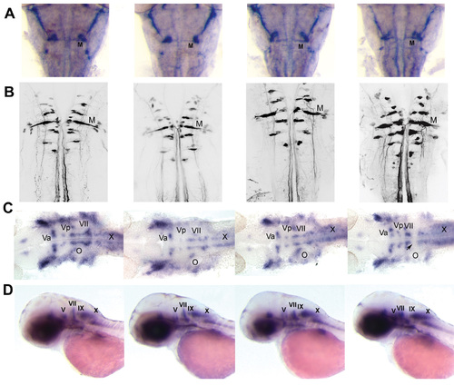
Patterns of primary and secondary hindbrain motorneurons in miR-10 overexpression and morphant embryos
Column 1; wildtype, column 2; morpholino 1 injected, column 3; morpholino 2 injected, column 4; miR-10 siRNA injected (also shown in main figure 7) A) 3A10 neurofilament immunolabeling. Mauthner neurons are present in morphant and overexpression embryos and are indistinguishable from wildtypes. B) Confocal images of hindbrains of 5 day old embryos retrograde labeled embryos. No differences are observed between non injected, morphant or overexpression embryos. C) Flatmounts of 30 hpf embryos in situ hybridized with islet-1. Patterns of branchiomotorneurons are indicated. Note the failure of the VIIth cranial nerve to migrate into r 5/6. The motorneuron patterns in the morphant embryos are like wildtypes. D) Sideview of 48 hpf embryos in situ hybridized with islet-1. In miR-10 siRNA injected embryos the VIIth nerve is located near the Vth nerve and there is a large gap between the VIIth and the IXth nerve. The patterns in the morphant embryos are like wildtypes.
|
