
FIGURE
Fig. 4
- ID
- ZDB-FIG-080723-3
- Publication
- Boisset et al., 2008 - Characterization of pip5k3 fleck corneal dystrophy-linked gene in zebrafish
- Other Figures
- All Figure Page
- Back to All Figure Page
Fig. 4
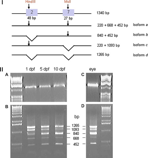
Spliced variants analysis during development. (I) Experimental procedure. Restriction site for HindIII and MslI in exon 2 and 7 are shown (arrows). Length of each fragments found after digestion for all isoforms are shown on the right. (II, A and C) Larval cDNA and adult eye cDNA before digestion. (II, B) Digested larval cDNA at stage 1 dpf, 3 dpf and 5 dpf showed a steady state expression of isoform a (668 bp) and isoform d (1265 bp). Isoform b (840 bp) is upregulated and Isoform c (1093 bp) is downregulated. (II, D) Isoforms a, b, c and d are present in adult eye, isoform b and d at high level. |
Expression Data
| Gene: | |
|---|---|
| Fish: | |
| Anatomical Terms: | |
| Stage Range: | Prim-5 to Adult |
Expression Detail
Antibody Labeling
Phenotype Data
Phenotype Detail
Acknowledgments
This image is the copyrighted work of the attributed author or publisher, and
ZFIN has permission only to display this image to its users.
Additional permissions should be obtained from the applicable author or publisher of the image.
Reprinted from Gene expression patterns : GEP, 8(6), Boisset, G., Polok, B.K., and Schorderet, D.F., Characterization of pip5k3 fleck corneal dystrophy-linked gene in zebrafish, 404-410, Copyright (2008) with permission from Elsevier. Full text @ Gene Expr. Patterns