
Fig. 4
- ID
- ZDB-IMAGE-080723-3
- Genes
- Publication
- Boisset et al., 2008 - Characterization of pip5k3 fleck corneal dystrophy-linked gene in zebrafish
- All Figures
- Figures for Boisset et al., 2008
|
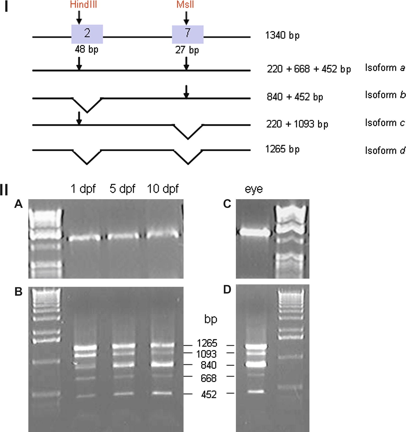
Fig. 4 Spliced variants analysis during development. (I) Experimental procedure. Restriction site for HindIII and MslI in exon 2 and 7 are shown (arrows). Length of each fragments found after digestion for all isoforms are shown on the right. (II, A and C) Larval cDNA and adult eye cDNA before digestion. (II, B) Digested larval cDNA at stage 1 dpf, 3 dpf and 5 dpf showed a steady state expression of isoform a (668 bp) and isoform d (1265 bp). Isoform b (840 bp) is upregulated and Isoform c (1093 bp) is downregulated. (II, D) Isoforms a, b, c and d are present in adult eye, isoform b and d at high level.
Reprinted from Gene expression patterns : GEP, 8(6), Boisset, G., Polok, B.K., and Schorderet, D.F., Characterization of pip5k3 fleck corneal dystrophy-linked gene in zebrafish, 404-410, Copyright (2008) with permission from Elsevier. Full text @ Gene Expr. Patterns