|
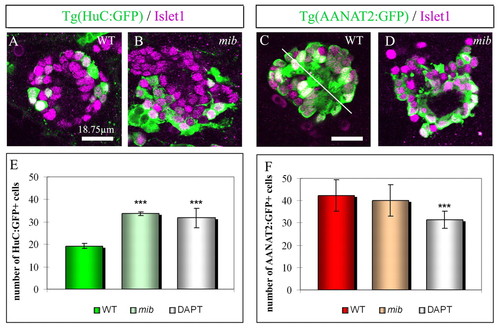
Modification of neuronal subtype identity in mib and DAPT-treated embryos. (A,B) Expression of GFP (green) and Islet1 (purple) in wild-type (WT) and mib;Tg(HuC:GFP) transgenic embryos at 48 hours. As Tg(HuC:GFP) labels other structures close to the epiphysis and as the epithalamus of mib embryos is highly disorganized, Islet1 serves to identify epiphysial neurons. (C,D) Expression of GFP (green) in Tg(AANAT2:GFP) transgenic embryos shown as confocal sections with Islet1 (purple). In wild-type embryos, Tg(AANAT2:GFP)+ photoreceptors are arranged in two mirror-imaged rows with the outer segments of the cells located at the midline (white line), this stereotyped organization is lost in mib embryos. Scale bars: 18.75 μm. (E,F) Average numbers of GFP+ cells (green) in Tg(HuC:GFP) (E) or in Tg(AANAT2:GFP) embryos (F) in the epiphysis of wild-type, mib or DAPT-treated embryos at 48 hours. Anterior is upwards. Error bars represent the standard deviation *P<0.05; ***P<0.0005 using a t-test.
|