
Fig. 7
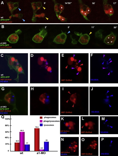
Phagolysosomal Fusion Defects in atp6v0a1 Morpholino Knock-Down (A) Confocal time-lapse analysis of wild-type (A) and atp6v0a1-MO microglia (B) microglia in 3 dpf brains. Time is indicated in minutes and seconds. Microglia are in green marked by GFP expression (Apo-E-GFP) while acidic endocytotic vesicles are marked in red by the LysoTracker. Blue arrowheads mark the formation of phagosomes. Yellow arrowheads mark phagosomal acidification (the vesicle turns red). White asterisks label single acidic vesicles. Fusion between acidic phagosomes is only observed in wild-type. (C–P) Heterotypic vesicular fusion in wild-type (C–F) and in atp6v0a1-MO microglia (G–P). (E, I, L, and O) NBT-DsRed labeling of phagosomes and phagolysosomes. (F, J, M, and P) DQ-BSA labeling of lysosomes and phagolysosomes. (D, H, K, and N). Merge. Phagosomes are marked by red arrowhead, lysosomes by blue arrowheads and phagolysosomes by purple arrowheads. Phagolysosomes (purple) are only observed in the wild-type (C and D). (Q) Percentage of the number of phagosomes (red bars), lysosomes (blue bars) and phagolysosomes (purple bars) present in wild-type (n = 15) and morphant cells (n = 15). |
| Fish: | |
|---|---|
| Knockdown Reagent: | |
| Observed In: | |
| Stage: | Protruding-mouth |
Reprinted from Cell, 133(5), Peri, F., and Nüsslein-Volhard, C., Live Imaging of Neuronal Degradation by Microglia Reveals a Role for v0-ATPase a1 in Phagosomal Fusion In Vivo, 916-927, Copyright (2008) with permission from Elsevier. Full text @ Cell