
Fig. 2
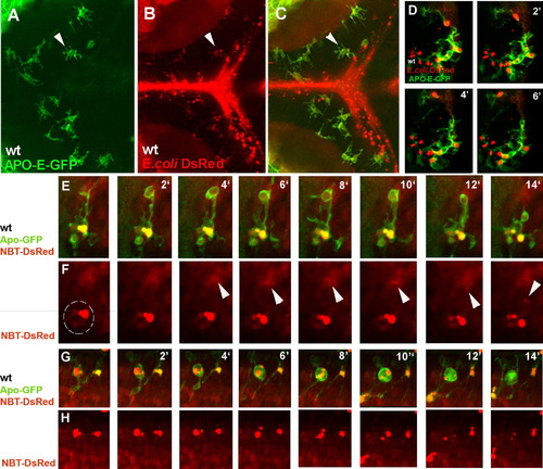
Microglia Phagocytose E. coli Bacteria and Neurons in the Brain (A–C) Dorsal views of a 3 dpf (days post fertilization) embryonic wild-type brain. (A) Microglial labeled by Apo-E-GFP. (B) Red-labeled Gram-negative E.coli bacteria (DsRed Express, Clontech). (C) Merge. Red bacterial inclusions are present inside the microglia (white arrowhead). (D) Confocal time-lapse of one Apo-E-GFP wild-type microglial cell collecting a red-labeled bacterial cluster (white arrowheads). (E–H) Confocal time-lapse of one branching Apo-E-GFP wild-type microglial cell collecting (E) and (F) and digesting (G) and (H) NBT-DsRed neurons. Time is indicated in minutes. White arrowheads point to the neuronal material being collected by a phagosome, the dotted line encircles the neuronal material inside the microglia (F). |
| Genes: | |
|---|---|
| Fish: | |
| Condition: | |
| Anatomical Terms: | |
| Stage: | Protruding-mouth |
Reprinted from Cell, 133(5), Peri, F., and Nüsslein-Volhard, C., Live Imaging of Neuronal Degradation by Microglia Reveals a Role for v0-ATPase a1 in Phagosomal Fusion In Vivo, 916-927, Copyright (2008) with permission from Elsevier. Full text @ Cell