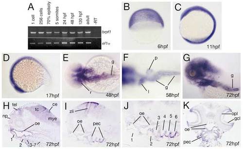
brpf1 displays early ubiquitous and later restricted expression in neuroectodermal, ectodermal and endodermal derivatives. (A) RT-PCR at indicated developmental stages for brpf1 and, as control, ef1α transcripts (for details, see legend to Fig. S1D). brpf1 is expressed at all stages investigated, from the 1-cell stage throughout adulthood (1-cell=0 hpf; 256-cell=early blastula stage=3 hpf; 70% epiboly=mid-gastrula stage=8 hpf; 5-somites=early segmentation stage=12 hpf). Transcripts detected during the first 4 hours of development, before the onset of zygotic transcription (Kane and Kimmel, 1993), are most likely maternally provided. To knockdown both maternal and zygotic brpf1 transcripts, we injected an antisense MO targeting the translational start site (in contrast to the described splice MO, which only targets zygotic transcripts). Preliminary results indicate that defects in segmental identity in such maternal-zygotic morphants are no more severe than in brpf1 mutants, with a regular initiation of anterior Hox gene expression (K.L. and M.H., unpublished; compare with Fig. S2). (B-K) brpf1 whole-mount in situ hybridization at stages indicated in lower right corners (6 hpf=shield stage=early gastrula; 11 hpf=3-somite stage; 17 hpf=16-somite stage). (B-D,G) Lateral views; (E,F) dorsal views; (H-J) longitudinal sections, (H) medial, (I,J) lateral; (K) horizontal section. Numbers of arches in H-J are indicated. ce, cerebellum; gcl, ganglion cell layer (retina); g, gut; l, liver; mye, myencephalon; np, nasal pit; oe, oral ectoderm; opl, outer plexiform layer (retina); pec, pharyngeal ectoderm; p, pancreas; r, posterior retina; tel, telencephalon; zli, zona limitans intrathalamica (Scholpp et al., 2006).
|
