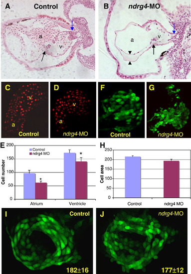
Attenuation of Ndrg4 results in smaller cardiac chambers and a reduction in the number of cardiomyocytes. Transverse sections of 3 dpf wild-type (A) and ndrg4 morphant (B) heart. Outflow tract (blue arrows), AV junction (black arrows) and the enlarged space (arrowheads) between myocardium and endocardium in the dilated atrium are indicated. Note only a few blood cells in the MO treated heart. Confocal images of the heart of control (C) and ndrg4-MO2 injected (D) Tg(cmlc2:DsRed2-nuc) embryos at 2 dpf. (E) Comparison of the numbers of cardiomyocytes in un-injected and ndrg4-MO2 injected embryos at 2 dpf (Data are from 5 control and 5 injected embryos and are expressed as means ± S.D. *P < 0.05). Confocal images of the heart of control (F) and ndrg4-MO2 injected (G) Tg(cmlc2:gfp) embryos at 2 dpf. (H) Comparison of the size of cardiomyocytes in control and ndrg4-MO2 injected embryos at 2 dpf. There was no change in myocardial cell number between control (I) and ndrg4-MO2 (J) injected Tg(cmlc2:gfp) embryos at 24 hpf based on the number of GFP positive cells in the heart: 182 ± 16 vs. 177 ± 12, respectively (Data are from 5 embryos for each group and are expressed as means ± S.D.) a, atrium; v, ventricle; control, mismatch-MO.
|
