Appl1-Mediated Gain-of-Function Phenotypes and Survival Signaling Depend on Its Endosomal Localization
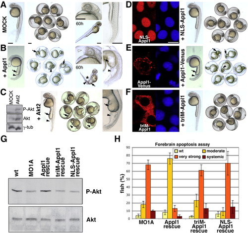
(A) Morphology of control embryos, injected with buffer only.
(B) Morphology of embryos injected with 500 pg appl1 mRNA. Note the thick yolk extension (arrows), blown up blood island (black arrowheads), and swollen telencephalon (white arrowheads).
(C) Western blot analysis and morphology of embryos overexpressing Akt2. Total and P-Akt levels are elevated in embryos that have been injected with 300 pg akt2 mRNA. γ-tubulin (γ-tub) was used as a loading control. The embryo on the left has received 300 pg mRNA, others 600 pg. Note thick yolk extensions (arrows) and blown up blood island (black arrowheads).
(D) Appl1 fused to a Nuclear Localization Signal (NLS-Appl1) is targeted to the nucleus and does not evoke phenotypes.
(E) Appl1 fused to a bulky tag (Appl1-Venus) is excluded from the nucleus and causes phenotypes identical to the one evoked by overexpression of WT Appl1 (B).
(F) Appl1 carrying the R147A, K153A, K155A triple mutation (triM-Appl1) fails to associate with endosomes and does not evoke phenotypes.
(A–F) Embryos after 26 hr of development, unless labeled otherwise. The scale bars (in [A]) represent 250 μm.
(D–F) Localization of three engineered Appl1 proteins and morphology of embryos injected with 500 pg of mRNA, respectively. Anti-Appl1 (red) and nuclear draq5 staining (blue). The scale bar (in [D]) represents 10 μm.
(G) Evaluation of Akt activity in WT, Appl1 knockdown (MO1A: 8 ng MO1A), and embryos in which a rescue with the WT appl1 mRNA (Appl1 rescue: 8 ng MO1A + 75 pg appl1 mRNA lacking MO1A binding site), the soluble triM-Appl1 (triM-Appl1 rescue: 8 ng MO1A + 75 pg triM-appl1 mRNA lacking MO1A binding site), or the nuclear NLS-Appl1 mutant (NLS-Appl1 rescue: 8 ng MO1A + 75 pg NLS-appl1 mRNA lacking MO1A binding site) has been attempted.
(H) Forebrain apoptosis assay: Quantitative comparison of apoptosis in embryos manipulated as in (G). Embryos were assigned to one of the four indicated cell death categories. Evaluation of Appl1 knockdown and WT appl1 mRNA–rescued embryos (Figure 4G), are shown again for comparison. Total amount of embryos scored: ntriM-Appl1 rescue = 567, nNLS-Appl1 rescue = 502. Error bars indicate SD amongst five experiments.
(G and H) Experiments have been carried out on 54 hr zebrafish embryos.
|

