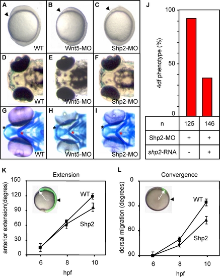
Shp2-MO–Induced CE Cell Movement Defects
Zebrafish embryos were not injected (A, D, G) or microinjected with Wnt5-MO (5 ng) (B, E, H) or Shp2-MO (1.0 ng) (C, F, I) at the one-cell stage.
(A–C) Morphology at 10 hpf shows reduced anterior extension of the Wnt5-MO– and Shp2-MO–injected embryos. Arrowheads indicate the anterior of the embryos
(D–F) Morphology of the Wnt5 and Shp2 knockdown embryos at 4 dpf show a mild hammerhead-like phenotype.
(G–I) Alcian blue staining of the cartilage in the heads of 4 dpf embryos. Black asterisk, Meckel′s cartilage; red asterisk, ceratohyal. (J) Zebrafish embryos were (co-) injected with Shp2-MO (1.0 ng) and 300 pg human shp2 mRNA and scored at 4 dpf.
(K,L) Embryos were loaded with caged fluorescein dextran and the fluorophore was uncaged at the shield stage (6 hpf) dorsally to determine anterior extension (K, white arrow in inset; site of uncaging, black arrowhead) or laterally to determine dorsal migration (L, white arrow in inset; initial position at the shield stage, black arrowhead). Cell labeling of the same embryos was followed immediately after uncaging at 80% epiboly (8 hpf) and at tailbud stage (10–10.5 hpf). WT and Shp2-MO–injected embryos were assessed and averages for ten embryos are given in degrees.
|
