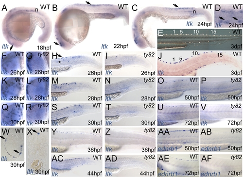
Expression pattern of zebrafish ltk in WT (A–D,F,H,J,K,M,O,Q,S,U,W–Y,AC) and shdty82 homozygous embryos (G,I,L,N,P,R,T,V,Z,AD) throughout embryonic development.
Stages indicated in hpf. A–C) ltk-expressing cells in vicinity of eye (lower arrows in A,B) and in premigratory trunk NC (upper arrow in B and C) and in notochord (n). D) Dorsal view of posterior trunk of WT embryo to show ltk expression in scattered cells in dorsolaterally-positioned subset of premigratory NCCs (arrows). E,J) WT embryo treated with phenylthiourea, illuminated with incident light to show iridophore pattern (E), then fixed and processed for ltk ISH (J); individual cells are numbered. F,G,K,L,Q,R) Dorsoventral spread of ltk-expression in WT eye (F,K,Q); cells remain dorsal to eye in shdty82 mutants (G,L,R). H,I,M,N,S,T,Y,Z,AC,AD) Cells in premigratory (arrow) and migratory (*) positions and in nascent dorsal and ventral stripes are prominent in WT (H,M,S,Y,AC), but almost absent from shdty82 mutants (I,N,T,Z,AD). O,P,U,V,AA,AB,AE,AF) ltk expression pattern closely resembles ednrb1 expression in WT iridophores (AA,AE), but both markers are absent in shdty82 homozygous embryos (P,V,AB,AF). W) Plastic section through eye. X) Transverse section of posterior trunk.
|
