Fig. 5
- ID
- ZDB-FIG-080331-7
- Publication
- Lee et al., 2008 - Olfactomedin-2 mediates development of the anterior central nervous system and head structures in zebrafish
- Other Figures
- All Figure Page
- Back to All Figure Page
|
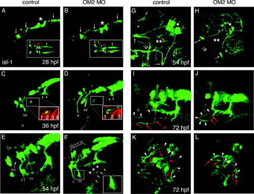
Axon guidance of branchiomotor neurons is perturbed in OM2 morphants. Head is to the left in A–F. All images were captured by a laser confocal microscope and z-stacked images were collapsed. (A and B) Lateral and dorsal view of 28 hpf embryos showing branchiomotor nuclei and initial axon outgrowth (n = 23 for A, n = 40 for B). Arrows show newly formed branchiomotor axon bundles and primary branchiomotor neurons. No apparent defects were detected at 28 hpf, except for fusion of the trigeminal nuclei in OM2 morphants (asterisks). (C and D) Embryos (36 hpf), showing normal development of branchiomotor nuclei in morphants (D), but mildly impaired outgrowth of branchiomotor axons (n = 20 for C, n = 44 for D). Inset in D shows failure of axons to grow into the pharyngeal arches in OM2 morphants (red: zn5 staining). Arrowheads in (C) indicate newly formed gill motor axons. Numbers in insets of (C and D) show pharyngeal endodermal pouches, absence/fusion of pouches in morphants. (E–H) Embryos (54 hpf) analyzed for branchiomotor development (n = 25 for E and G, n = 73 for F and H). In control islet-1-GFP fish (E), cranial nerve IV, V and VII are labeled with Roman numerals. SoL indicates superior orbital lateral line axons. (F) Lateral view of branchiomotor axons of a 54 hpf ATG OM2 morphant fish. Islet-1-derived GFP expression shows the stereotypically perturbed projection of cranial nerve V and VII with abrupt endings and numerous fine filopodia (inset in F). Gill motor neurons from CN X do not form nerve bundles that innervate gill muscles (asterisks in F). Despite the relatively normal position of branchiomotor nuclei V and VII, close observation reveals slightly hypomorphic nuclei in morphants (bracket with CN V and VII in F). Aberrant axons with anteriorily wandering processes were detected from the vagus (X) nerve, as indicated by arrows in (F). Absence of SoL is indicated by red asterisks. (G) Ventral view of 54 hpf control fish. The characteristic crossed axon pathway formed by cranial nerve V and VII is apparent (double arrow). Single arrows indicate intermediate branches of cranial nerve V. (H) In OM2 ATG morphants cranial nerve V and VII prematurely terminate as shown in (F), which results in the absence of the crossed axon pathway (double arrow). Notice also the absence of peripherally extended cranial nerve VII in morphants (double asterisk). Single asterisk indicates hypomorphic telencephalic nuclei. (I) Lateral view of 72 hpf control fish. Anteriorily projected CN V and VII are indicated by white arrowheads (V) and red arrows (VII). (J) Lateral view of 72 hpf OM2 morphant fish. One of the well-recovered fish is shown here. CN V axon (white arrowheads) grew further compared with (F) and made a stereotypic medio-caudal turn, whereas CN VII axons did not grow much further (red arrows). (K) Ventral view of 72 hpf control fish. As in (G) a cross-shaped axon pathway made by CN V and CN VII axons is readily visible (white arrowheads and red arrows). (L) Ventral view of 72 hpf OM2 morphant. White arrowheads indicate the axon pathway formed by further-projected CN V axons, which could reach the midline in this case. Notice much more caudal location of the axon pathway compared with that in control fish (K). Red arrows indicate CN VII axons that did not grow further from the earlier observation (54 hpf ventral view, H). |
| Fish: | |
|---|---|
| Knockdown Reagent: | |
| Observed In: | |
| Stage Range: | Prim-5 to Long-pec |
Reprinted from Mechanisms of Development, 125(1-2), Lee, J.A., Anholt, R.R., and Cole, G.J., Olfactomedin-2 mediates development of the anterior central nervous system and head structures in zebrafish, 167-181, Copyright (2008) with permission from Elsevier. Full text @ Mech. Dev.

