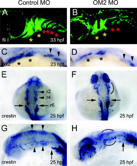
Analysis of cranial neural crest cells in OM2 morphants. (A and B) Confocal micrographs of 33 hpf fliI-GFP fish. Head is left. The first and second (yellow) asterisks indicate the developing mandibular and hyoid arches, which are only mildly affected by OM2 knockdown. More caudal pharyngeal arches (red asterisks) are severely affected in OM2 morphants. (C and D) dlx2 in situ hybridization of 23 hpf fish. Asterisks in (C) and (D) indicate the migrated cranial neural crest cells within the developing mandibular and hyoid arches. Cranial neural crest cells in more caudal pharyngeal arches are indicated by arrowheads. Although hypomorphic, the pattern and position of the dlx2-positive cells suggest that initial specification and migration of cranial neural crest cells are normal in OM2 morphants. (E–H) Crestin in situ hybridization of 25 hpf zebrafish. Rostral streams of cranial neural crest cell migration are indicated by r2, r4 and r6, which originate from rhombomeres 2, 4 and 6, respectively. In morphants (F and H), r2 and r4 streams were not well segregated (68/70: dotted lines) and r6 stream was hypomorphic (69/70).
|
